Searching…
    Home Product Ultrasound Systems B/W Ultrasound Systems Sonoview

    Sonoview

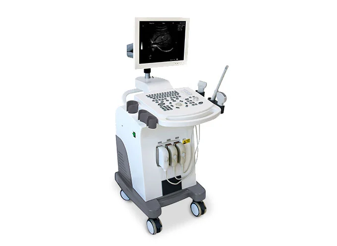
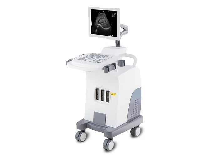
    Fully digital 3D-ready ultrasound with broadband probes and mobile trolley.

    SONOVIEW® Ultrasound Diagnostic System Core Information

    1‌.Host Configuration‌: Fully digital imaging platform equipped with a 15-inch color LCD display, supports PC-based extended functionalities, and is upgradeable to 3D imaging.

    2.Probe System‌: Broadband multi-frequency probes delivering high-clarity imaging.

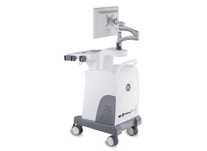
    ‌3.Supporting Facilities‌: Dedicated mobile trolley.

    CE CE Marked
    ABS Made by ABS
    LATEX Not made with natural rubber latex

    Product Features

    Discover the advanced capabilities that set our products apart

    SONOVIEW® Ultrasound System Core Specifications (Refined)
    1. Display & Imaging Performance‌

    Screen‌: 15-inch color LCD with pseudo-color display (8 types) and adjustable image direction.
    Resolution‌: Lateral 2mm, Longitudinal 1mm; Scanning depth up to 240mm.
    Image Quality‌: Fully digital technology ensuring high clarity; 256 grayscale levels.
    Processing‌: Advanced pre/post-processing, dynamic range adjustment, edge enhancement, inversion, and γ-correction.
    2. Probe & Scanning Capabilities‌

    Standard Probe‌: 3.5 MHz electronic convex array (5-segment frequency conversion).
    Optional Probes‌: 7.5 MHz linear, 6.5 MHz intracavity (transvaginal), 7.5 MHz rectal, and 3.5 MHz micro-convex.
    Focusing‌: 4 electronic foci with random combination.
    Modes‌: B, 2B, B/M, M, 4B, ZOOM (real-time in B mode), and optional 3D imaging.
    3. Data Management & Functions‌

    Storage‌: High-capacity local memory + USB support; Cine-Loop recording (user-preset: 64–1024 images).
    Software‌: PC-based architecture with full application packages, advanced measurement/calculation, and powerful reporting/management tools.
    Connectivity‌: Compatible with laser/inkjet printers; optional R/W DVD-ROM.
    4. Configuration Summary‌

    Standard‌: Mainframe (15" LCD) + 3.5 MHz convex probe.
    Options‌: Additional probes (linear, intracavity, rectal, micro-convex), DVD-ROM, and laser printer.

    Product Specifications

    Detailed technical specifications and performance parameters

    Display depth: 240mm Resolution: lateral 2mm, longitudinal 1mm Probe multi-frequency conversion: 5 segment frequencies TGC setting: 8 TGC settings Grayscale: 256 Scanning and display modes: B, 2B, B/M, M, 4B, ZOOM; real-time zoom in B mode; 3D (optional) Main probe: 3.electronic convex 5 MHz array (frequency conversion) Electronic focus: 4 foci in random combination Image processing: pre-processing, post-processing, dynamic range, frame rate, line average, edge; enhancement, black/white inversion; grayscale adjustment, contrast, brightness, γ-revision. Image direction: top/bottom/left/right Pseudo color function: 8 types Magnification: ratio 10, 1.5, 2.0, 2.5, 3.0, 3.5, 4.0, 4.5, 5.0, 5.5, 6.0 Cine loop: 64, 128, 256, 512, 1024 frames (preset by user) Body markers: a variety of body markers (30 types) Information display: Date, time, medical record number, magnification, measurement value, body marker, character hints, image correlation coefficient, scan depth, probe type portfolio, conversion to English and Chinese, full-screen character editing, etc. Measurement and calculation: B mode: distance, circumstance, area, volume, angle, ratio, stenosis, profile, histogram; M mode: heart rate, time, distance, slope and stenosis; Gynecologic measurements: Uterus, cervix, endometrium, ovary L/R; Obstetrics: gestational age, fetal weight, AFI; Cardiology: LV, LV function, LVPW, RVAWT; Urology: volume of the transition zone, bladder volume, RUV, prostate, kidney; Small parts: Optics, thyroid, jaw and face. DICOM3.0: Medical Digital Imaging and Communication, is the industry standard (agreement) for image and data transmission between different types of medical devices. Ultrasound devices can receive images and data via DICOM when connected to a PACS. System preset: System preset includes parameter presets for gynecology, gynecology, vascular, cardiology, urology and small parts, comments, manufacturing default settings, system upgrade and maintenance settings. Memory: large capacity of local memory and USB memory, image and cine loop, measurement result and report can be saved. Monitor: 14" black and white SVGA display with progressive scan Probe connection: 2 Terminal output: VGA, PAL-D video output Size: 790mm (length) × 660mm (width) × 1070mm (height) Weight: approx. 50kg

    Related Accessories

    Compatible accessories to extend functionality

    Standard:
    • Mobile trolley   .............................. 1Pcs
    • Multi frequency convex probe ...... 1Pcs
    • Power Cable  ............................... 1Pcs
    • Cleaning Cloth  ............................ 1Pcs
     
    Options:
    • 7.5MHz high-frequency linear array probe
    • 6.5MHz intra-cavity (trans-vaginal) probe
    • 7.5MHz rectal probe
    • 3.5MHz micro-convex probe
    • R/W DVD-ROM
    • B/W Laser printer

    Product Overview

    Core specifications at a glance

    SKU MDT17-D00004
    Gross Weight 38kg
    Package Size 83x63x95cm
    QTY/BOX 1pcs